Home > Popular Themes > Human Body
Bladder epithelium, light micrograph

![]()
Wall Art and Photo Gifts from Science Photo Library
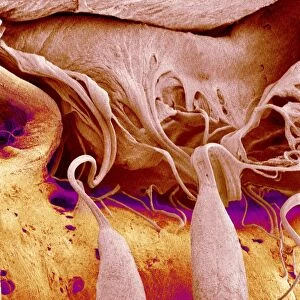
Bladder epithelium, light micrograph
Bladder epithelium. Light micrograph of a vertical section through the wall of the urinary bladder. The inner surface is at top. The upper layer (light pink) is the transitional epithelium, which is especially adapted for the bladders function of containing urine. The plasma membranes surrounding the epithelial cells here are much thicker than most cell membranes, with a specialised sub-structure. The epithelium is thus rendered impermeable to potentially toxic urine. Below the epithelium is smooth muscle and connective tissue (dark red). Magnification: x3300 when printed 10 centimetres wide
Science Photo Library features Science and Medical images including photos and illustrations
Media ID 6450253
© STEVE GSCHMEISSNER/SCIENCE PHOTO LIBRARY
Adaptation Adapted Bladder Connective Tissue Epithelial Epithelium Histological Histology Internal Surface Longitudinal Plasma Membrane Smooth Muscle Specialised Thickened Tissue Urinary System Urology Wall Cells Light Micrograph Light Microscope Section Sectioned Thicker
EDITORS COMMENTS
This print showcases the intricate structure of bladder epithelium, providing a glimpse into the remarkable adaptation of this vital organ. The image reveals a vertical section through the wall of the urinary bladder, with its inner surface displayed at the top. The upper layer, depicted in a delicate shade of pink, represents the transitional epithelium. This specialized tissue plays a crucial role in maintaining the bladder's function by containing urine. What sets it apart is its unique cellular composition - plasma membranes surrounding these epithelial cells are noticeably thicker than typical cell membranes and possess a specialized sub-structure. This ingenious design renders the epithelium impermeable to potentially harmful substances present in urine. Beneath this protective layer lies smooth muscle and connective tissue, portrayed in deep shades of red. These underlying structures provide support and contribute to proper bladder function. With an impressive magnification level of x3300 when printed 10 centimeters wide, this light micrograph offers an awe-inspiring view into our internal anatomy. It serves as a testament to both the complexity and resilience of our bodies. This stunning visual representation not only highlights normal anatomical features but also provides valuable insights for urology specialists studying various conditions affecting urinary system health.
MADE IN THE USA
Safe Shipping with 30 Day Money Back Guarantee
FREE PERSONALISATION*
We are proud to offer a range of customisation features including Personalised Captions, Color Filters and Picture Zoom Tools
SECURE PAYMENTS
We happily accept a wide range of payment options so you can pay for the things you need in the way that is most convenient for you
* Options may vary by product and licensing agreement. Zoomed Pictures can be adjusted in the Cart.