Stressed cells

![]()
Wall Art and Photo Gifts from Science Photo Library
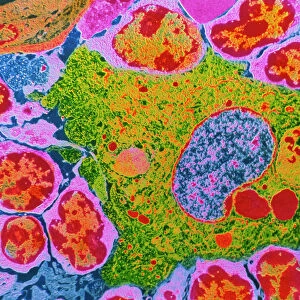
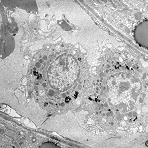
Stressed cells
Stressed cells (image 2 of 2). Immunofluorescent light micrograph of stressed kidney cells. Stress is caused by chemicals, UV light, viral infection and heat shock. The cell enters an emergency mode. TIA-1 and TIAR proteins stop the production (translation) of all proteins, except those needed during stress. The two proteins also assist in the formation of stress granules (blue-green dots), cytoplasmic material where untranslated genetic material (mRNA) can be stored. During these processes, the cell has time to decide whether to make more proteins or shut down activity. The nuclei of the cells (green) and a new protein, FAST K (red), are shown
Science Photo Library features Science and Medical images including photos and illustrations
Media ID 6450017
© NANCY KEDERSHA/SCIENCE PHOTO LIBRARY
Cytological Cytology Granule Immunofluorescence Kidney Messenger Ribonucleic Acid Microtubules Mrna Nuclei Nucleus Physiological Physiology Proteins Translation Light Micrograph Light Microscope Protein
EDITORS COMMENTS
This print showcases the intricate world of stressed kidney cells, revealing the fascinating mechanisms they employ to cope with various stressors. The image, captured using immunofluorescent light microscopy, vividly illustrates how chemicals, UV light, viral infection, and heat shock trigger a cellular emergency response. In this state of distress, two crucial proteins called TIA-1 and TIAR step in as saviors. They halt the production of most proteins within the cell except for those essential during times of stress. Moreover, these remarkable proteins also aid in forming stress granules - depicted as mesmerizing blue-green dots scattered throughout the cytoplasm. These granules serve as repositories where untranslated genetic material (mRNA) can be stored temporarily. Amidst these intricate processes unfolding within the cell's interior, it gains precious time to evaluate whether it should continue protein synthesis or cease its activities altogether. The nuclei of these resilient cells are highlighted in vibrant green hues while a newly discovered protein known as FAST K is represented by striking red tones. This awe-inspiring micrograph not only offers valuable insights into animal biology but also delves into translation physiology at a cellular level. It explores the dynamic interplay between nucleus and cytoplasm while unraveling the critical roles played by mRNA and various proteins such as TIA-1 and TIAR during moments of intense cellular stress.
MADE IN THE USA
Safe Shipping with 30 Day Money Back Guarantee
FREE PERSONALISATION*
We are proud to offer a range of customisation features including Personalised Captions, Color Filters and Picture Zoom Tools
SECURE PAYMENTS
We happily accept a wide range of payment options so you can pay for the things you need in the way that is most convenient for you
* Options may vary by product and licensing agreement. Zoomed Pictures can be adjusted in the Cart.